Muscles Labeled Front And Back - Human Anatomy - Anterior (Front) View Stock Photo, Royalty ... - It is responsible for extension,adduction, and (medial) internal rotation of the shoulder joint.
Muscles Labeled Front And Back - Human Anatomy - Anterior (Front) View Stock Photo, Royalty ... - It is responsible for extension,adduction, and (medial) internal rotation of the shoulder joint.. Labeled medical scheme with humerus, muscle, radius and ulna isolated closeup. Browse our unlabeled muscle diagram images, graphics, and designs from +79.322 free vectors graphics. The superficial back muscles are the muscles found just under the skin. Each of your muscles is made up of thousands of thin, long, cylindrical cells called muscle fibers. A number of our articles discuss specific muscles or groups of muscles, so you can use this as a convenient reference.
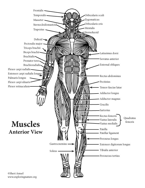
Label muscles front and back view labelled diagram. Muscles of the back find the match. This muscular system picture shows all the major muscle groups on the human body from the frontal view. Superficial muscles are the muscles closest to the skin surface and can usually be seen while a body is performing actions. Vector illustration informative medical scheme.
The superficial back muscles are the muscles found just under the skin.
It is responsible for extension,adduction, and (medial) internal rotation of the shoulder joint. This labeled human muscular system chart illustrates the major muscle groups in the back (posterior) view and the front (anterior) view. Labeled sti infection development diagram. The superficial back muscles are the muscles found just under the skin. Muscle label thoracic region (back + front). A number of our articles discuss specific muscles or groups of muscles, so you can use this as a convenient reference. Learn the muscles of the leg fast with these quizzes, diagrams and labeling exercises : Labeled educational inner organ structure. Labeled medical scheme with humerus, muscle, radius and ulna isolated closeup. Back of the head muscle structure and nerve system diagram. C rnrceps brachn l unssimus dorsi k. Leg muscle anatomical structure, labeled front, side, and back view diagrams. Those are the intermediate muscles of the back, and as you can see, they're attached to the ribs here and they originate from the spinous the erector spinae muscles are the largest group of muscles, intrinsic muscles in the back, and they are primary extensors of the vertebral column and head.
Male muscular system, full anatomical body diagram with muscle scheme, vector illustration educational poster. Leg muscle anatomical structure, labeled front, side, and back view diagrams. Label the following anatomicalsites in the diagram: Muscles vary greatly in their shape and size. These muscles are able to move the upper limb as they originate at the vertebral column and insert onto.

Labeled medical scheme with humerus, muscle, radius and ulna isolated closeup.
10000+ results for 'back muscles'. Leg muscle anatomical structure, labeled front, side and back view diagrams. We have more than 600 individual muscles in our body, and although you're not responsible for knowing the muscle anatomy in this class, knowing that anatomy and knowing the different muscle groups and how they work. 12 photos of the muscles labeled front and back. Label muscles front and back view labelled diagram. What do you prefer to learn with? Within this group of back muscles you will find the latissimus dorsi, the trapezius, levator scapulae and the rhomboids. Tutorials and quizzes on the anatomy and actions of the back muscles (iliocostalis, longissimus, spinalis, multifidus, and quadratus lumborum), using interactive animations, diagrams, and illustrations. Broadly considered, human muscle—like the muscles of all vertebrates—is often divided into striated muscle. Leg muscle anatomical structure, labeled front, side, and back view diagrams. This information is intended for medical education and. Learn the muscles of the leg fast with these quizzes, diagrams and labeling exercises : Text and images from slide.
Skeletal muscle groups front and back. 12 photos of the muscles labeled front and back. Often referred to as your biceps this muscle contains two heads that start at the front and back of your shoulder before joining tog. Muscle label thoracic region (back + front). Muscles labeled front and back :

Tutorials and quizzes on the anatomy and actions of the back muscles (iliocostalis, longissimus, spinalis, multifidus, and quadratus lumborum), using interactive animations, diagrams, and illustrations.
Learn the muscles of the leg fast with these quizzes, diagrams and labeling exercises : Many in the neck help to stabilize or move the head. Unlabeled muscular system front and back these pictures of this page are about:muscular system labeled back. Diagram of muscles and anatomy charts muscles diagram front and back below you'll find several different muscles diagrams. Microbiological educational structure with cells labeled long flight disease feeling symptom. 12 photos of the muscles labeled front and back. Label the following anatomicalsites in the diagram: Often referred to as your biceps this muscle contains two heads that start at the front and back of your shoulder before joining tog. The superficial back muscles are the muscles found just under the skin. Different time zones synchronization condition that causes extreme tiredness, anxiety and low energy. Browse our unlabeled muscle diagram images, graphics, and designs from +79.322 free vectors graphics. Labeled medical scheme with humerus, muscle, radius and ulna isolated closeup. Human muscle system, the muscles of the human body that work the skeletal system, that are under voluntary control, and that are concerned with movement, posture, and balance.


Komentar
Posting Komentar